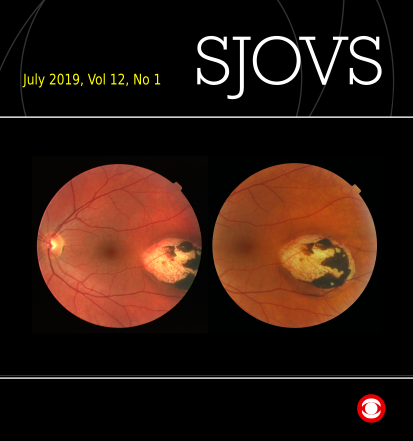
Ocular toxoplasmosis with surprisingly good retinal function
DOI:
https://doi.org/10.5384/sjovs.vol12i1p1-4Keywords:
Ocular toxoplasmosis, retinal function, OCT, scotoma, visual field, retinochoroidal scarAbstract
Ocular toxoplasmosis is an infection in the eye caused by the parasite Toxoplasma Gondii. A common retinal finding in its inactive stages are pigmented retinochoroidal scarring. The retinal function in the affected area assumingly reflects the amount of retinal involvement. This case report presents a 48-year-old woman who has a long-standing large retinochoroidal scar in the temporal posterior pole of her left eye. She had not experienced any visual symptoms, and no recurrent infections had occurred as far as she knew. She has a scotoma in her nasal visual field that her optometrist detected by a coincidence when she was in her twenties. The corresponding visual field defect is smaller and less deep than what may be expected from the structural appearance of the scar. The reported case demonstrates, that the visual function may be well preserved in the visual field corresponding to a retinochoroidal scarred area due to toxoplasmosis, in spite of loss of structures in the outer retinal layers as seen with OCT.
Metrics
References
Bosch-Driessen, L. H., Karimi, S., Stilma, J. S., & Rothova, A. (2000). Retinal detachment in ocular toxoplasmosis. Ophthalmology, 107(1), 36-40.
Cabral, C. M., Tuladhar, S., Dietrich, H. K., Nguyen, E., MacDonald, W. R., Trivedi, T., . . . Koshy, A. A. (2016). Neurons are the Primary Target Cell for the Brain-Tropic Intracellular Parasite Toxoplasma gondii. PLOS Pathogens, 12(2), e1005447. doi:10.1371/journal.ppat.1005447
Cassels, N. K., Wild, J. M., Margrain, T. H., Chong, V., & Acton, J. H. (2018). The use of microperimetry in assessing visual function in age-related macular degeneration. Surv Ophthalmol, 63(1), 40-55. doi:10.1016/j.survophthal.2017.05.007
Commodaro, A. G., Belfort, R. N., Rizzo, L. V., Muccioli, C., Silveira, C., Burnier Jr, M. N., & Belfort Jr, R. (2009). Ocular toxoplasmosis: an update and review of the literature. Mem Inst Oswaldo Cruz, 104(2), 345-350.
Furtado, J. M., Winthrop, K. L., Butler, N. J., & Smith, J. R. (2013). Ocular toxoplasmosis I: parasitology, epidemiology and public health. Clin Exp Ophthalmol, 41(1), 82-94. doi:10.1111/j.1442-9071.2012.02821.x
Garg, S., Mets, M. B., Bearelly, S., & Mets, R. (2009). Imaging of congenital toxoplasmosis macular scars with optical coherence tomography. Retina, 29(5), 631-637. doi:10.1097/IAE.0b013e318198d8de
Gilbert, R. E., Dunn, D. T., Lightman, S., Murray, P. I., Pavesio, C. E., Gormley, P. D., . . . Stanford, M. R. (1999). Incidence of symptomatic toxoplasma eye disease: aetiology and public health implications. Epidemiology and Infection, 123(2), 283-289.
Goldenberg, D., Goldstein, M., Loewenstein, A., & Habot-Wilner, Z. (2013). Vitreal, retinal, and choroidal findings in active and scarred toxoplasmosis lesions: a prospective study by spectral-domain optical coherence tomography. Graefe's Archive for Clinical and Experimental Ophthalmology, 251(8), 2037-2045. doi:10.1007/s00417-013-2334-3
Harrell, M., & Carvounis, P. E. (2014). Current Treatment of Toxoplasma Retinochoroiditis: An Evidence-Based Review. Journal of Ophthalmology, 2014, 7. doi:10.1155/2014/273506
Hildebrand, G. D., & Fielder, A. R. (2011). Anatomy and Physiology of the Retina. In J. D. Reynolds & S. E. Olitsky (Eds.), Pediatric retina (pp. 39-65). Heidelberg: Springer.
Holland, G. N. (2009). Ocular toxoplasmosis: the influence of patient age. Mem Inst Oswaldo Cruz, 104(2), 351-357.
Jonas, J. B., & Panda-Jonas, S. (2016). Secondary Bruch's membrane defects and scleral staphyloma in toxoplasmosis. Acta Ophthalmol, 94(7), e664-e666. doi:10.1111/aos.13027
Lavinsky, D., Romano, A., Muccioli, C., & Belfort, R., Jr. (2012). Imaging in ocular toxoplasmosis. Int Ophthalmol Clin, 52(4), 131-143. doi:10.1097/IIO.0b013e318265fd78
Meenken, C., Assies, J., van Nieuwenhuizen, O., Holwerda-van der Maat, W. G., van Schooneveld, M. J., Delleman, W. J., . . . Rothova, A. (1995). Long term ocular and neurological involvement in severe congenital toxoplasmosis. Br J Ophthalmol, 79(6), 581-584.
Ng, P., & McCluskey, P. J. (2002). Treatment of Ocular Toxoplasmosis. Australian Prescriber, 25(4), 88-90.
Padhi, T. R., Das, S., Sharma, S., Rath, S., Rath, S., Tripathy, D., . . . Besirli, C. G. (2017). Ocular parasitoses: A comprehensive review. Surv Ophthalmol, 62(2), 161-189. doi:https://doi.org/10.1016/j.survophthal.2016.09.005
Park, Y.-H., & Nam, H.-W. (2013). Clinical Features and Treatment of Ocular Toxoplasmosis. The Korean Journal of Parasitology, 51(4), 393-399. doi:10.3347/kjp.2013.51.4.393
Pleyer, U., Schluter, D., & Manz, M. (2014). Ocular toxoplasmosis: recent aspects of pathophysiology and clinical implications. Ophthalmic Res, 52(3), 116-123. doi:10.1159/000363141
Rensch, F., & Jonas, J. B. (2008). Direct microperimetry of alpha zone and beta zone parapapillary atrophy. Br J Ophthalmol, 92(12), 1617-1619. doi:10.1136/bjo.2008.139030
Scherrer, J., Iliev, M. E., Halberstadt, M., Kodjikian, L., & Garweg, J. G. (2007). Visual function in human ocular toxoplasmosis. Br J Ophthalmol, 91(2), 233-236. doi:10.1136/bjo.2006.100925
Stanford, M. R., Tomlin, E. A., Comyn, O., Holland, K., & Pavesio, C. (2005). The visual field in toxoplasmic retinochoroiditis. Br J Ophthalmol, 89(7), 812-814. doi:10.1136/bjo.2004.055756
Sutton, M. S., & Torbit, J. K. (2001). Toxoplasmosis. In K. H. Thomann, E. S. Marks, & D. T. Adamczyk (Eds.), Primary eyecare in systemic disease (2nd ed., pp. 571-581). USA: McGraw-Hill, Medical Pub. Div.
Downloads
Published
How to Cite
Issue
Section
License
Authors retain copyright and grant the journal right of first publication with the work simultaneously licensed under a Creative Commons Attribution License that allows others to share the work with an acknowledgement of the work's authorship and initial publication in this journal.


